Laboratory work number 8 biology. Practical and laboratory work in biology (grade 8). Products necessary for its preparation
Amankaragai secondary school named after N. Ostrovsky

Laboratory work in biology
8th grade
(deep study)
A guide for teachers and students


Compiled by Mazhara E.G.,
biology teacher
Amankaragai
Laboratory work number 1.Subject: Carrying out anthropological measurements: height, weight, establishing correlations between the sizes of individual parts of the body. Purpose: to establish the relationship of changes in indicators of physical developmentperson with age.Equipment: centimeter tape, stadiometer. Progress.1. Height measurement Height is measured using a stadiometer. The subject must stand on the platform of the stadiometer, touching the vertical stand with the heels, buttocks, interscapular region and the back of the head. The experimenter measures the growth of the subject and records the result. 2. Measurement circles chest cells Experimenter using measuring tape measures the circumference chest. To do this, the subject raises his hands, the experimenter applies the tape so that it passes along the lower corners of the shoulder blades. In front, the tape should pass along the mid-sternal point and fit snugly to the body. Then the subject lowers his hands. The circumference of the chest is measured in three phases: during normal calm breathing (in a pause), with maximum inhalation and maximum exhalation. Determine the excursion of the chest - the difference between the values of the circumference of the chest on exhalation and inhalation. Record the result. 3. Determination of body weight The measurement is carried out using medical scales. 4. The results obtained are drawn up in the following form:Observation progress:
test subject 5. Make a conclusion about the change in indicators of physical developmentperson with age.
Laboratory work number 2.
Topic: The study of the structure of cells and tissues of the human body under a microscope.
Purpose: to study the structure of cells and tissues of the human body under a microscope.
Equipment: table "Structure of cells and organelles", textbook.
Progress.
1
. Consider the drawing. Fill in the corresponding rows of the table "Cage":
2. Fill in the table:
Structural system of the nucleus
structures
3. Compare diagrams of the distribution of elements in the earth's crust and their content in living organisms. Why are the most common elements in nature, except for oxygen, in living organisms are presented in very small quantities (less than 0.1%)?
R 
Distribution of elements in the earth's crust (A) and in living organisms (B)
4. Make a conclusion about the structure of the cell of the human body.
Laboratory work number 3.
Subject: The study of the knee jerk and the observation of the knee jerk during the experiment.
Target: observation of the occurrence of a knee jerk under mechanical action.Equipment: hammer from a children's designer.Progress:conduct an experiment: the first student, the subject, in a sitting position on a chair, puts his right foot on his left. The second student, the experimenter, inflicts a light blow with a hammer on the tendon of the muscle of the right leg (knee joint). The experiment is repeated with the left leg.
Compare the reflex response to mechanical action.
Make a conclusion.
Laboratory work number 4.
Topic: Physiological tests illustrating the work of the cerebellum.
Target: introduce students to the functions of the cerebellum.Equipment:Progress:1. Finger-nose test
The subject closes his eyes, stretches his right hand forward with a straightened index finger, the remaining fingers are clenched into a fist. After that, touch your nose with the tip of your index finger.Evaluation of results Normally, a healthy person performs this task. If the function of the cerebellum is impaired, this task is feasible only if the hand is lowered down.
2. Braking of movements arising due to inertia
The work is done in pairs. The subject bends his arm at the elbow. The experimenter grabs his forearm near the hand and invites the subject to pull his hand towards himself, overcoming the resistance. Then, unexpectedly for the subject, the experimenter releases his hand. The subject's hand makes a short jerk and stops.3. Draw conclusions by answering the following questions.- What function of the cerebellum did you determine using a finger-nose test? What function of the cerebellum did you determine with the help of inhibition of movements that arose due to inertia? Why, when an intoxicated person tries to take one step, he often takes several steps in the same direction by inertia?
Lab #5
Topic: Unconditioned reflexes of the medulla oblongata, midbrain and diencephalon.
Target:
get acquainted with the unconditioned reflexes of the medulla oblongata, midbrain and diencephalon.Equipment: table "The structure of the brain."Progress
1. Medulla oblongata
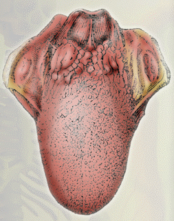
With the handle of a spoon, the experimenter touches the back surface of the tongue. Involuntarily there is a swallowing reflex.The subject makes several swallowing movements in a row. When there is no saliva left in his mouth, the swallowing reflex will not appear.The subject takes 2-3 quick and deep breaths and exhalations. After that, his breathing stops for a while..
- What functions of the medulla oblongata were revealed in these experiments? What other functions of this part of the brain are familiar to you?
- What functions of the midbrain have been established with the help of these experiments? You must have noticed that in public places
doors most often open outward - what function of the midbrain is this associated with?
- What are the reflexes whose centers are located in the diencephalon, hypothalamus? What are the functions of the hypothalamus in the diencephalon?
Laboratory work number 6.
Subject:Determination of visual acuity.
Target: determine visual acuity with the help of experiments.Equipment: frames measuring 15x20 cm with well-stretched gauze, a set of objects of different colors.Progress:
- Break up in pairs. One student places a frame with stretched gauze in front of his eyes at a distance of 29 cm, behind which, at a distance of 50 cm, another student places a page of the textbook. The first student, on command, fixes his gaze first on the gauze threads, then on the text. The experience is repeated several times. As a result, students are convinced that it is impossible to see letters and a gauze pattern at the same time. One student sits on a chair and looks straight ahead. Another student alternately demonstrates a set of objects painted in different colors. The subject is shown in motion and for a short time. Each demonstration should be accompanied by questions: What item was shown? What colour? Draw your own conclusions.
Laboratory work number 7.
Subject:Determination of hearing acuity.
Target: determine hearing acuity empirically.Equipment: table "Structure of the organs of hearing", centimeter tape.Progress:
- Consider the figure and table “The structure of the hearing organs. Break up in pairs. One student at a distance of 10 cm reads the text from the textbook in a low voice, then the distance increases and the distance at which the student stops hearing is recorded in the notebook. Then they change places. the teacher turns on the music player and changes the volume of the sound. The pitch of the perceived sound is determined. Draw your own conclusions.
- Try bending and then stretching the animal's natural bone. Did she bend over? Were you able to stretch it? What happens when you try to bend a calcined bone? What properties does it have? Is it possible to stretch a bone in hydrochloric acid? What properties does this bone have?
Laboratory work number 9.
Subject:Providing first aid for sprains, dislocations and fractures of bones.
Target: learn how to provide first aid for injuries.Equipment: tires, bandage, gauze napkins, scarf.Progress:
Learn how to put on a pressure bandage. When is it applied?
Provide first aid for a fracture of the forearm, shoulder, lower leg, thigh.
The victim has a fracture of the bones of the skull, another spine, chest. Provide first aid.
Make a conclusion.
Laboratory work number 10.
Topic: Determination of the location of bones and muscles during external examination.
Target:
locate bones and muscles.Equipment: tables, figures.Progress:
1. Consider the diagrams of the skeletal and muscular systems.2. Fill in the table
3. Draw conclusions by answering the following questions.
- What provides a certain body shape? How are muscles fixed? Why is it possible for individual parts of the body to move relative to each other? What muscles flex and extend the human hand? Where are the muscles that flex the fingers located? What muscle lifts the heel? What movement is involved in the deltoid muscle? What muscles flex and extend the leg knee joint?
What muscles allow you to maintain a vertical position of the body?
Laboratory work number 11.
Subject:Anthropometric method for determining the level of growth and development of the body
Target: learn to measure and evaluate indicators of physical development. Equipment: stadiometer, floor scales, centimeter tape.
Progress:
1. Height measurement
Height is measured using a stadiometer. The subject must stand on the platform of the stadiometer, touching the vertical stand with the heels, buttocks, interscapular region and the back of the head. The experimenter measures the growth of the subject and records the result.
2. Measurement circles chest cells
The experimenter uses a measuring tape to measure the circumference of the chest. To do this, the subject raises his hands, the experimenter applies the tape so that it passes along the lower corners of the shoulder blades. In front, the tape should pass along the mid-sternal point and fit snugly to the body. Then the subject lowers his hands. The circumference of the chest is measured in three phases: during normal calm breathing (in a pause), with maximum inhalation and maximum exhalation.
Determine the excursion of the chest - the difference between the values of the circumference of the chest on exhalation and inhalation. Record the result.
3. Determination of body weight
The measurement is carried out using medical scales.
Note: The study should be conducted on at least 5 subjects of different ages (preschooler, schoolchild, adult).
The results obtained are presented in the following form:
test subject
4. Draw a conclusion about the change in indicators of physical development of a person with age.
Laboratory work number 12.
Topic: Microscopic structure of human and frog blood.
Target:
compare the structure of human and frog blood cells.Equipment: microscope, frog and human blood microslides.Progress:
- Examine a sample of human blood under a microscope. Find red blood cells and draw them. Consider a micropreparation of frog blood. Sketch the red blood cells of a frog. Find the differences between human and frog erythrocytes. Answer the question: Whose blood carries more oxygen - the blood of a person or a frog. Why? Make a conclusion about the difference in the structure of human and frog blood, using the data in the table:
Laboratory work number 13.
Subject:Immunity. AIDS prevention.
Target: learn to distinguish between types of immunity.Equipment: textbook, drawings.Progress:
1. Answer the following questions in writing:
What is immunity, what are the types of immunity?
Which cells in the body are responsible for immune responses?
How is immunity different in children?
What is immunization and why is it carried out?
2.Fill in the table:
Types of immunity
3. Make a conclusion.Laboratory work No. 14.Subject: External and internal structure of the heart. Target: to study the features of the external and internal structure hearts. Equipment: table, drawings. Progress.
- Look at the picture and answer the question: Where is the heart located?

2. Look at the picture. Sketch the internal structure of the heart (Fig. 80) and designate the structural components:

3. Draw a conclusion, answering the question: What devices in the structure of the heart ensure the movement of blood in one direction?
Lab #15Subject: Self-observation. functional tests. Target: get acquainted with functional tests that allow you to find out the degree of fitness of your heart. Equipment: stopwatch. Progress: 1. Measure your resting heart rate. To do this, take 3-4 measurements for
10 s and multiply the average value by 6.
2. Do 20 squats at a fast pace, sit down and immediately measure your heart rate for 10 seconds.
3. Repeat measurements every 20 seconds. Determine the heart rate for 10 s. (For measurements, 20 s is counted from the end of the previous measurement.)
4. Present your results in the form of a table below. It contains approximate values that may not match yours.
Heart rate at rest
5. Draw a conclusion.
Note: The results are good if the heart rate after squats increased by 1/3 or less of the resting results; if half - the results are average, and if more than half - the results are unsatisfactory.
Evaluation of results The pulse rate at the age of 15-20 years is normally 60-90 beats per minute. In the supine position, the pulse is on average 10 beats per minute less than in the standing position. In women, the pulse is 7-10 beats per minute more often than in men of the same age. The pulse rate during work in the range of 100 - 130 beats per minute indicates a low intensity of the load. The frequency of 130 - 150 beats per minute characterizes the load of medium intensity. Frequency 150 - 170 beats per minute - the load is above average intensity. The frequency of 170 - 200 beats per minute is characteristic of the maximum load.Lab #16Subject: Measurement of blood pressure before and after dosed exercise. Target: learn how to measure blood pressure. Equipment: tonometer. Progress: 1. Measure your blood pressure with a blood pressure monitor. Compare the data obtained in the experiment with the average table data on blood pressure for your age. Make a conclusion.2. Calculate the values of pulse (PP), mean arterial (APm) and intrinsic arterial pressure (APsyst and APdiast). It is known that normal healthy person pulse pressure is approximately 45 mm Hg. arterial (BP): ADsyst. = 1.7 x age + 83
ADdiast. = 1.6 x age + 42 Pulse (PD): PD = ADsist. - ADdiast. Mean arterial (APav): Addr. \u003d (BP system - AD diast.) / 3 + AD diast.Evaluation of results Compare the calculated data obtained in the experiment with the data presented in the table.

3. Make a conclusion, p by answering the questions:
- What is the danger to a person is constantly high pressure? In what vessels of our body is the lowest pressure and why?
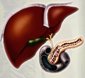
- Could the solution enter the left ventricle?
2) Where could he penetrate if it is known that the entrance to the coronary artery is located in the wall of the aorta and is covered by semilunar valves during the ejection of blood?
3) Why was adrenaline included in the solution in addition to nutrients and oxygen?
4) What feature of the heart muscle made it possible to revive the heart outside the body?
Lab #17
Subject:Providing first aid for bleeding.
Target: learn to recognize types of bleeding, provide first aid for damage to blood vessels.Equipment: bandage, tourniquet, napkins.Progress:
1. The victim has severe bleeding from a wound on his right forearm, the blood is coming in jerks, the color of the blood is scarlet. Render first aid and explain what kind of bleeding it is. Provide first aid. Explain.3. The victim has an abrasion on his knee, the bleeding is weak, the wound is dirty. Provide first aid. Explain. 4. Draw a conclusion. Lab #18
Subject:Methods for measuring the frequency and depth of respiratory movements.
Target: establish the effect of holding the breath on the respiratory rate.Equipment: stopwatch (watch with a second hand).Progress:
1. Determine the time to hold your breath while inhaling in a sitting position. The subject breathes calmly for 3-4 minutes in a sitting position, and then, on command, after a normal exhalation, takes a deep breath and holds his breath as long as he can, while holding his nose. The experimenter, using a stopwatch, determines the time from the moment of holding the breath to the moment of its resumption. The result is fixed. 2. Do 20 squats in 30 seconds and again determine the breath holding time on inspiration.
3. Rest exactly 1 minute and repeat step 1.Evaluation of results 4. Evaluate the results using the table

5. Draw a conclusion.
Lab #19
Topic: Study of the action of saliva enzymes on starch.
Target:
make sure that there are enzymes in saliva that can break down starch.Equipment:
a piece of starched dry bandage the size of a palm, a Petri dish or a saucer with a weak solution of iodine, cotton swabs.Progress:
1. Moisten a Q-tip with saliva and write a letter in the middle of a piece of starched bandage. 2. Hold the gauze between your palms for 2-3 minutes, and then lower it into the iodine solution.
3. Watch how the piece of gauze stains.4. Draw a conclusion about what happened and why.
Laboratory work number 20.
Topic: Compiling a diet for a teenager.
Target: learn how to make a daily diet for teenagers.
Equipment: tables chemical composition food products and calories, daily energy needs of children and adolescents of different ages, daily norms of proteins, fats and carbohydrates in the food of children and adolescents.
Progress:
When compiling a human diet, the following rules should be followed:– the calorie content of the diet should correspond to the daily energy consumption;- it is necessary to take into account the optimal amount of proteins, fats and carbohydrates for persons engaged in these types of labor (and for children - age);
- the best diet involves four meals a day (first breakfast should be 10-15%, second breakfast - 15-35%, lunch - 40 - 50% and dinner 15-20% of the total calories);
- Protein-rich foods (meat, fish, eggs) are more rational to use for breakfast and lunch. For dinner, you should leave dairy and vegetable dishes;
- in the diet, about 30% should be proteins and fats of animal origin.With a mixed diet, a person digests an average of about 90% of food.
1. Make a daily diet for a teenager 15-16 years old
2. Enter the result of the calculations in the table.
3. Draw conclusions:- on the calorie content of the diet, on the optimality of the diet, on the fulfillment of daily norms in the consumption of nutrients.
The composition of the daily diet
Diet
General conclusions:
The calorie content of the diet should correspond to the daily energy consumption.
When choosing the optimal diet, it is important to take into account not only the calorie content, but also the chemical components of food.
It is necessary to take into account the ratio of proteins, fats and carbohydrates in the diet, their characteristics in food products of various origins.
Calculation of energy costs and determination of the calorie content of the diet.
Calculations can be carried out after performing any physical activity. The formula allows you to set the energy consumption committed by a person in 1 minute, according to the heart rate (HR). The formula for calculating the energy consumption of a person in 1 min for any physical activityQ = 2.09(0.2 x HR–11.3) kJ/minExample . Let's say you skied for 30 minutes, your heart rate reached 120 beats per minute. Let's calculate the energy consumption for 1 minute:Q \u003d 2.09 (0.2 x 120 - 11.3) \u003d 2.09 (24 - 11.3) \u003d 26.5 kJ / min.Answer : 795 kJ was consumed in 30 minutes.Calculate the energy expenditure of a person who swam in the pool for 15 minutes, after which the heart rate reached 130 beats per minute.Based on the result, draw a conclusion about the dependence of the amount of energy expended on the heart rate.The composition of food products and their calorie content
The product's name
Daily norms of proteins, fats and carbohydrates in the food of children and adolescents
Age, years
Daily energy requirement of children and adolescents of different ages (kcal)
Age, years
Lab #21
Subject:External and internal structure of the kidney
Target:
to study the features of the external and internal structure of the human kidney.Equipment: textbook, table “Structure of excretory organs, micropreparation, microscope.Progress:
- Examine a micropreparation of a kidney under a microscope. Fill in the tables using the textbook material:
excretory system.
Organs
Stages of urination
- Make a conclusion.
Lab #22
Subject:Examination of the dorsal and palmar surfaces of the hand.
Target:
compare the structure of the dorsal and palmar sides of the hand.Equipment: drawings.Progress:
- Examine the back and palmar side of the hand. Compare the skin on the dorsal and palmar sides. Consider the location of the skin folds, drawings. Make a conclusion about the features of the dorsal and palmar sides of the hand.
Amankaragai secondary school named after N. Ostrovsky
Laboratory work in biology
8th grade
(deep study)
A guide for teachers and students
Compiled by Mazhara E.G.,
biology teacher
Amankaragai
Laboratory work number 1.
Subject: Carrying out anthropological measurements: height, weight, establishing correlations between the sizes of individual parts of the body.
Purpose: to establish the relationship of changes in indicators of physical development
person with age.
Equipment: centimeter tape, stadiometer.
Progress.
1. Height measurement
Height is measured using a stadiometer. The subject must stand up.
on the platform of the stadiometer, touching the vertical stand with the heels, buttocks,
interscapular region and occiput. Experimenter measures height
subject and write down the result.
2. Measurement circles chest cells
An experimenter using a centimeter tape measures the circumference of the chest
cells. To do this, the subject raises his hands, the experimenter imposes
tape so that it runs along the lower corners of the shoulder blades. The front tape should
pass along the mid-sternal point and fit snugly to the body. Then
the subject lowers his hands. Chest circumference is measured in three phases: during
normal calm breathing (in a pause), with maximum inspiration and maximum
exhale. Determine the excursion of the chest - the difference between the values
chest circumference on exhalation and inhalation. Record the result.
3. Determination of body weight
The measurement is carried out using medical scales.
4. The results obtained are drawn up in the following form:
Observation progress:
test subject
Height
chest circumference
Body mass
5. Make a conclusion about the change in indicators of physical development
person with age.
Laboratory work number 2.
Topic: The study of the structure of cells and tissues of the human body under a microscope.
Purpose: to study the structure of cells and tissues of the human body under a microscope.
Equipment: table "Structure of cells and organelles", textbook.
Progress.
1
. Consider the drawing. Fill in the corresponding rows of the table "Cage":
The name of the components and organelles of the cell
Structure
2. Fill in the table:
Structural system of the nucleus
structures
Structure
3. Compare diagrams of the distribution of elements in the earth's crust and their content in living organisms. Why are the most common elements in nature, except for oxygen, in living organisms are presented in very small quantities (less than 0.1%)?
R
Distribution of elements in the earth's crust (A) and in living organisms (B)
4. Make a conclusion about the structure of the cell of the human body.
Laboratory work number 3.
Subject: The study of the knee jerk and the observation of the knee jerk during the experiment.
Target: observation of the occurrence of a knee jerk under mechanical action.
Equipment: hammer from a children's designer.
Progress:
conduct an experiment: the first student, the subject, in a sitting position on a chair, puts his right foot on his left. The second student, the experimenter, inflicts a light blow with a hammer on the tendon of the muscle of the right leg (knee joint). The experiment is repeated with the left leg.
Compare the reflex response to mechanical action.
Make a conclusion.
Laboratory work number 4.
Topic: Physiological tests illustrating the work of the cerebellum.
Target: introduce students to the functions of the cerebellum.
Equipment:
Progress:
1. Finger-nose test
The subject closes his eyes, stretches his right hand forward with a straightened index finger, the remaining fingers are clenched into a fist. After that, touch your nose with the tip of your index finger.
Evaluation of results
Normally, a healthy person performs this task. If the function of the cerebellum is impaired, this task is feasible only if the hand is lowered down.
2. Braking of movements arising due to inertia
The work is done in pairs. The subject bends his arm at the elbow. The experimenter grabs his forearm near the hand and invites the subject to pull his hand towards himself, overcoming the resistance. Then, unexpectedly for the subject, the experimenter releases his hand. The subject's hand makes a short jerk and stops.
What function of the cerebellum did you determine using a finger-nose test?
What function of the cerebellum did you determine with the help of inhibition of movements that arose due to inertia?
Why, when an intoxicated person tries to take one step, he often takes several steps in the same direction by inertia?
Lab #5
Topic: Unconditioned reflexes of the medulla oblongata, midbrain and diencephalon.
Target: get acquainted with the unconditioned reflexes of the medulla oblongata, midbrain and diencephalon.
Equipment: table "The structure of the brain."
Progress
1. Medulla oblongata
With the handle of a spoon, the experimenter touches the back surface of the tongue. Involuntarily there is a swallowing reflex.
The subject makes several swallowing movements in a row. When there is no saliva left in his mouth, the swallowing reflex will not appear.
The subject takes 2-3 quick and deep breaths and exhalations. After that, his breathing stops for a while.
.
What functions of the medulla oblongata were revealed in these experiments?
What other functions of this part of the brain are familiar to you?
2. Midbrain
The experimenter offers the subjects tasks (for example, read a short text). As soon as all the subjects began to read, he suddenly and quite forcefully taps on the table with a pencil. At this point, most subjects will stop reading and involuntarily turn their head towards the source of the sound (orienting reflex).
The subject looks at a lit lamp. One light source is visible. Now he gently presses down on one of the eyeballs and looks back at the light source. The object begins to double, two light bulbs are visible. This happened because the correct setting of the eye, controlled by the midbrain, was disturbed.
The subject closes his eyes, stretches his right hand forward with the index finger extended, the remaining fingers are clenched into a fist. After that, touch your nose with the tip of your index finger.
Answer the following questions.
What functions of the midbrain have been established with the help of these experiments?
You must have noticed that in public places doors most often open outward - what function of the midbrain is this associated with?
3. Diencephalon
The experimenter invites the subjects to mind their own business. And then suddenly gives a loud command: "Freeze!". The subjects freeze in different positions (late diencephalon reflex).
4. Draw conclusions by answering the following questions:
What are the reflexes whose centers are located in the diencephalon, hypothalamus?
What are the functions of the hypothalamus in the diencephalon?
Laboratory work number 6.
Subject:Determination of visual acuity.
Target: determine visual acuity with the help of experiments.
Equipment: frames measuring 15x20 cm with well-stretched gauze, a set of objects of different colors.
Progress:
Break up in pairs. One student places a frame with stretched gauze in front of his eyes at a distance of 29 cm, behind which, at a distance of 50 cm, another student places a page of the textbook. The first student, on command, fixes his gaze first on the gauze threads, then on the text. The experience is repeated several times. As a result, students are convinced that it is impossible to see letters and a gauze pattern at the same time.
One student sits on a chair and looks straight ahead. Another student alternately demonstrates a set of objects painted in different colors. The subject is shown in motion and for a short time. Each demonstration should be accompanied by questions: What item was shown? What colour?
Draw your own conclusions.
Laboratory work number 7.
Subject:Determination of hearing acuity.
Target: determine hearing acuity empirically.
Equipment: table "Structure of the organs of hearing", centimeter tape.
Progress:
Consider the figure and table “The structure of the hearing organs.
Break up in pairs. One student at a distance of 10 cm reads the text from the textbook in a low voice, then the distance increases and the distance at which the student stops hearing is recorded in the notebook. Then they change places.
the teacher turns on the music player and changes the volume of the sound. The pitch of the perceived sound is determined.
Draw your own conclusions.
Laboratory work number 8.
Subject: Study of the properties of decalcified and calcined bones.
Target: to determine the difference between decalcified and calcined bones.
Progress:
Try bending and then stretching the animal's natural bone. Did she bend over? Were you able to stretch it?
What happens when you try to bend a calcined bone? What properties does it have?
Is it possible to stretch a bone in hydrochloric acid? What properties does this bone have?
Conclusion: What is the difference between decalcified and calcined bones?
Laboratory work number 9.
Subject:Providing first aid for sprains, dislocations and fractures of bones.
Target: learn how to provide first aid for injuries.
Equipment: tires, bandage, gauze napkins, scarf.
Progress:
Learn how to put on a pressure bandage. When is it applied?
Provide first aid for a fracture of the forearm, shoulder, lower leg, thigh.
The victim has a fracture of the bones of the skull, another spine, chest. Provide first aid.
Make a conclusion.
Laboratory work number 10.
Topic: Determination of the location of bones and muscles during external examination.
Target: locate bones and muscles.
Equipment: tables, figures.
Progress:
1. Consider the diagrams of the skeletal and muscular systems.
2. Fill in the table
3. Draw conclusions by answering the following questions.
What provides a certain body shape?
How are muscles fixed?
Why is it possible for individual parts of the body to move relative to each other?
What muscles flex and extend the human hand?
Where are the muscles that flex the fingers located?
What muscle lifts the heel?
What movement is involved in the deltoid muscle?
What muscles flex and extend the leg at the knee joint?
What muscles allow you to maintain a vertical position of the body?
Laboratory work number 11.
Subject:Anthropometric method for determining the level of growth and development of the body
Target: learn to measure and evaluate indicators of physical development.
Equipment: stadiometer, floor scales, centimeter tape.
Progress:
1. Height measurement
Height is measured using a stadiometer. The subject must stand on the platform of the stadiometer, touching the vertical stand with the heels, buttocks, interscapular region and the back of the head. The experimenter measures the growth of the subject and records the result.
2. Measurement circles chest cells
The experimenter uses a measuring tape to measure the circumference of the chest. To do this, the subject raises his hands, the experimenter applies the tape so that it passes along the lower corners of the shoulder blades. In front, the tape should pass along the mid-sternal point and fit snugly to the body. Then the subject lowers his hands. The circumference of the chest is measured in three phases: during normal calm breathing (in a pause), with maximum inhalation and maximum exhalation.
Determine the excursion of the chest - the difference between the values of the circumference of the chest on exhalation and inhalation. Record the result.
3. Determination of body weight
The measurement is carried out using medical scales.
Note: The study should be conducted on at least 5 subjects of different ages (preschooler, schoolchild, adult).
The results obtained are presented in the following form:
test subject
chest circumference
Body mass
4. Draw a conclusion about the change in indicators of physical development of a person with age.
Laboratory work number 12.
Topic: Microscopic structure of human and frog blood.
Target: compare the structure of human and frog blood cells.
Equipment: microscope, frog and human blood microslides.
Progress:
Examine a sample of human blood under a microscope. Find red blood cells and draw them.
Consider a micropreparation of frog blood. Sketch the red blood cells of a frog.
Find the differences between human and frog erythrocytes.
Answer the question: Whose blood carries more oxygen - the blood of a person or a frog. Why?
Make a conclusion about the difference in the structure of human and frog blood, using the data in the table:
Laboratory work number 13.
Subject:Immunity. AIDS prevention.
Target: learn to distinguish between types of immunity.
Equipment: textbook, drawings.
Progress:
1. Answer the following questions in writing:
What is immunity, what are the types of immunity?
Which cells in the body are responsible for immune responses?
How is immunity different in children?
What is immunization and why is it carried out?
2.Fill in the table:
Types of immunity
Congenital
Acquired
Action
Ways of formation
3. Make a conclusion.
Laboratory work No. 14.
Subject: External and internal structure of the heart.
Target: study the features of the external and internal structure of the heart.
Equipment: table, drawings.
Progress.
Look at the picture and answer the question: Where is the heart located?
What is the structure of the circulatory system?
2. Look at the picture. Sketch the internal structure of the heart (Fig. 80)
and label the structural components:
3. Draw a conclusion, answering the question: What devices in the structure of the heart ensure the movement of blood in one direction?
Lab #15
Subject: Self-observation. functional tests.
Target: get acquainted with functional tests that allow you to find out the degree of fitness of your heart.
Equipment: stopwatch.
Progress:
1. Measure your resting heart rate. To do this, take 3-4 measurements for
10 s and multiply the average value by 6.
2. Do 20 squats at a fast pace, sit down and immediately measure your heart rate for 10 seconds.
3. Repeat measurements every 20 seconds. Determine the heart rate for 10 s. (For measurements, 20 s is counted from the end of the previous measurement.)
4. Present your results in the form of a table below. It contains approximate values that may not match yours.
Heart rate at rest
Dynamics of heart rate recovery in seconds after work (20 squats)
10 s average
13
11
12
12
(13+11+12+12):4=12
5. Draw a conclusion.
Note: The results are good if the heart rate after squats increased by 1/3 or less of the resting results; if half - the results are average, and if more than half - the results are unsatisfactory.
Evaluation of results
The pulse rate at the age of 15-20 years is normally 60-90 beats per minute. In the supine position, the pulse is on average 10 beats per minute less than in the standing position. In women, the pulse is 7-10 beats per minute more often than in men of the same age. The pulse rate during work in the range of 100 - 130 beats per minute indicates a low intensity of the load. The frequency of 130 - 150 beats per minute characterizes the load of medium intensity. Frequency 150 - 170 beats per minute - the load is above average intensity. The frequency of 170 - 200 beats per minute is characteristic of the maximum load.
Lab #16
Subject: Measurement of blood pressure before and after dosed exercise.
Target: learn how to measure blood pressure.
Equipment: tonometer.
Progress:
1. Measure your blood pressure with a blood pressure monitor. Compare the data obtained in the experiment with the average table data on blood pressure for your age. Make a conclusion.
2. Calculate the values of pulse (PP), mean arterial (APm) and intrinsic arterial pressure (APsyst and APdiast). It is known that a normal pulse pressure in a healthy person is approximately 45 mm Hg. Art.
Arterial (BP):
ADsyst. = 1.7 x age + 83
ADdiast. = 1.6 x age + 42
Pulse (PD):
PD = ADsist. - ADdiast.
Mean arterial (APav):
Addr. \u003d (BP system - AD diast.) / 3 + AD diast.
Evaluation of results
Compare the calculated data obtained in the experiment with the data presented in the table.
3. Make a conclusion, p by answering the questions:
What is the danger to a person is constantly high pressure?
In what vessels of our body is the lowest pressure and why?
Exercise:
For the first time, the human heart was revived 20 hours after the death of a patient in 1902 by the Russian scientist Alexei Alexandrovich Kulyabko (1866-1930). The scientist sent a nutrient solution enriched with oxygen and containing adrenaline into the heart through the aorta.
Could the solution enter the left ventricle?
2) Where could he penetrate if it is known that the entrance to the coronary artery is located in the wall of the aorta and is covered by semilunar valves during the ejection of blood?
3) Why was adrenaline included in the solution in addition to nutrients and oxygen?
4) What feature of the heart muscle made it possible to revive the heart outside the body?
Lab #17
Subject:Providing first aid for bleeding.
Target: learn to recognize types of bleeding, provide first aid for damage to blood vessels.
Equipment: bandage, tourniquet, napkins.
Progress:
1. The victim has severe bleeding from a wound on his right forearm, the blood is coming in jerks,
the color of blood is scarlet. Give first aid and explain what kind of bleeding it is.
2. The victim has a skull injury: his forehead is cut, bleeding is profuse, the bone is not damaged. Provide first aid. Explain.
3. The victim has an abrasion on his knee, the bleeding is weak, the wound is dirty. Provide first aid. Explain.
4. Draw a conclusion.
Lab #18
Subject:Methods for measuring the frequency and depth of respiratory movements.
Target: establish the effect of holding the breath on the respiratory rate.
Equipment: stopwatch (watch with a second hand).
Progress:
1. Determine the time to hold your breath while inhaling in a sitting position. The subject breathes calmly for 3-4 minutes in a sitting position, and then, on command, after a normal exhalation, takes a deep breath and holds his breath as long as he can, while holding his nose. The experimenter, using a stopwatch, determines the time from the moment of holding the breath to the moment of its resumption. The result is fixed.
2. Do 20 squats in 30 seconds and again determine the breath holding time on inspiration.
3. Rest exactly 1 minute and repeat step 1.
Evaluation of results
5. Draw a conclusion.
Lab #19
Topic: Study of the action of saliva enzymes on starch.
Target: make sure that there are enzymes in saliva that can break down starch.
Equipment: a piece of starched dry bandage the size of a palm, a Petri dish or a saucer with a weak solution of iodine, cotton swabs.
Progress:
1. Moisten a Q-tip with saliva and write a letter in the middle of a piece of starched bandage.
2. Hold the gauze between your palms for 2-3 minutes, and then lower it into the iodine solution.
3. Watch how the piece of gauze stains.
4. Draw a conclusion about what happened and why.
Laboratory work number 20.
Topic: Compiling a diet for a teenager.
Target: learn how to make a daily diet for teenagers.
Equipment: tables of the chemical composition of food products and calorie content, daily energy needs of children and adolescents of different ages, daily norms of proteins, fats and carbohydrates in the food of children and adolescents.
Progress:
When compiling a human diet, the following rules should be followed:
– the calorie content of the diet should correspond to the daily energy consumption;
- it is necessary to take into account the optimal amount of proteins, fats and carbohydrates for persons engaged in these types of labor (and for children - age);
- the best diet involves four meals a day (first breakfast should be 10-15%, second breakfast - 15-35%, lunch - 40 - 50% and dinner 15-20% of the total calories);
- Protein-rich foods (meat, fish, eggs) are more rational to use for breakfast and lunch. For dinner, you should leave dairy and vegetable dishes;
- in the diet, about 30% should be proteins and fats of animal origin.
With a mixed diet, a person digests an average of about 90% of food.
1. Make a daily diet for a teenager 15-16 years old
2. Enter the result of the calculations in the table.
3. Draw conclusions:- on the calorie content of the diet, on the optimality of the diet, on the fulfillment of daily norms in the consumption of nutrients.
The composition of the daily diet
Diet
Name of the dish
Products necessary for its preparation
Weight, g
Calorie content, kcal.
Squirrels,
Fats
Carbohydrates
1st breakfast
2nd breakfast
Dinner
Dinner
General conclusions:
The calorie content of the diet should correspond to the daily energy consumption.
When choosing the optimal diet, it is important to take into account not only the calorie content, but also the chemical components of food.
It is necessary to take into account the ratio of proteins, fats and carbohydrates in the diet, their characteristics in food products of various origins.
Calculation of energy costs and determination of the calorie content of the diet.
Calculations can be carried out after performing any physical activity. The formula allows you to set the energy consumption committed by a person in 1 minute, according to the heart rate (HR). The formula for calculating the energy consumption of a person in 1 min for any physical activity
Q = 2.09(0.2 x HR–11.3) kJ/min
Example . Let's say you skied for 30 minutes, your heart rate reached 120 beats per minute. Let's calculate the energy consumption for 1 minute:
Q \u003d 2.09 (0.2 x 120 - 11.3) \u003d 2.09 (24 - 11.3) \u003d 26.5 kJ / min.
Answer : 795 kJ was consumed in 30 minutes.
Calculate the energy expenditure of a person who swam in the pool for 15 minutes, after which the heart rate reached 130 beats per minute.
Based on the result, draw a conclusion about the dependence of the amount of energy expended on the heart rate.
The composition of food products and their calorie content
The product's name
Squirrels
Fats
Carbohydrates
Calories per 100g. product, kcal.
in percentages
Buckwheat
12,5
67,4
351,5
Semolina
11,2
73,3
354,6
Rice
75,8
352,0
Pasta
11,0
74,2
358,4
Beans
23,2
53,8
355,7
Rye bread
42,9
222,6
wheat bread
47,0
234,6
Potato
20,0
90,2
Carrot
41,0
Beet
10,4
48,6
fresh cabbage
29,1
Sauerkraut
12,6
Green onion
23,3
watermelons
39,37
melons
11,3
49,8
fresh cucumbers
13,8
Pickles
6,92
Tomatoes
19,5
oranges
41,05
Grape
16,2
69,4
Lemons
10,3
44,6
tangerines
10,0
44,6
Apples
11,2
47,9
Rafinated sugar
99,9
41,7
Chocolate
37,2
53,2
59,7
Cocoa
23,6
20,2
40,2
450,3
Sunflower oil
99,8
930,3
Butter
83,5
782,3
Kefir
64,4
Sour cream
30,0
302,1
Curd
12,5
16,0
15,0
262,05
Fat cottage cheese
15,0
18,0
233,4
Creamy ice cream
10,0
17,0
179,4
Cheese
22,5
25,0
339,8
beef meat
20,0
10,7
181,8
lamb meat
19,0
132,9
Meat, lean pork
23,5
10,0
189,7
Goose
16,5
29,0
338,1
Chicken
20,0
128,6
Amateur sausage
13,7
27,9
316,2
sausages
12,4
19,4
233,4
Eggs
12,5
12,0
165,1
Salo
91,0
856,3
Bream
16,8
139,8
Zander
19,0
85,4
Cod
17,6
75,8
Red caviar
31,6
13,8
258,4
Herring
19,7
24,5
12,4
308,8
Eggplant caviar
13,0
158,9
Daily norms of proteins, fats and carbohydrates in the food of children and adolescents
Age, years
Proteins, g
Fats, g
Carbohydrates, g
50-60
60-70
150-200
65-70
75-80
250-300
8-11
75-95
80-95
350-400
12-14
90-110
90-110
400-500
15-16
100-120
90-110
450-500
Daily energy requirement of children and adolescents of different ages (kcal)
Age, years
Total based on average body weight
1603 – 1804
1804 – 2305
8-11
2355 – 2906
12-14
2806 –3307
15-16
3207 - 3508
Lab #21
Subject:External and internal structure of the kidney
Target: to study the features of the external and internal structure of the human kidney.
Equipment: textbook, table “Structure of excretory organs, micropreparation, microscope.
Progress:
Examine a micropreparation of a kidney under a microscope.
Fill in the tables using the textbook material:
excretory system.
Organs
Structure
Functions
Ureters
Urethra
Stages of urination
Where is happening
Components
Filtration
Reabsorption
Secretion
Make a conclusion.
Lab #22
Subject:Examination of the dorsal and palmar surfaces of the hand.
Target: compare the structure of the dorsal and palmar sides of the hand.
Equipment: drawings.
Progress:
Examine the back and palmar side of the hand.
Compare the skin on the dorsal and palmar sides.
Consider the location of the skin folds, drawings.
Make a conclusion about the features of the dorsal and palmar sides of the hand.
Laboratory works
to the course "Biology Grade 8"
LAB #1
on the topic: "Catalytic activity of enzymes"
Target: observe the catalytic function of enzymes in living cells.
Equipment: 1) 2 tubes
2) water bottle
3) raw and boiled potatoes
4) hydrogen peroxide (3%)
Progress:
1. Pour water into test tubes to a height of about 3 cm.
2. In one, add 3-4 pieces the size of a pea of raw potatoes, in the other - the same amount of boiled.
3. Pour 5-6 drops of hydrogen peroxide into each.
Formulation of results:
Describe what happened in the first and second test tubes. Sketch the experience.
What is the name of the substance that accelerates chemical reaction?
What is an enzyme? Under what conditions does it operate?
Doconclusion, explaining the results of the experiments.
LABORATORY WORK № 2
on the topic "Human tissues under a microscope"
Target: get acquainted with the microscopic structure of some tissues of the human body, learn how to identify them distinctive features
Equipment: 1) microscope
2) micropreparations:
* for option 1: "Glandular epithelium", "Hyaline cartilage",
* for option 2: "Nervous tissue", "Smooth muscles"
Progress:
Prepare the microscope for work and examine the micropreparations.
Formulation of results: Write down what you see in your notebook.
Doconclusion , listing the distinctive features of the tissues you saw (the type and location of cells, the shape of the nucleus, the presence of intercellular substance)
LABORATORY WORK № 3
on the topic: "Structure of bone tissue"
Target: to get acquainted with the structure of tubular and flat bones.
Equipment: 1) handout "Bone cuts"
2) sets of vertebrae
Progress:
1. Consider cuts of flat and tubular bones, find a spongy substance, consider its structure, in which bones is there a cavity? What is it for?
Formulation of results:
Sketch in your notebook what you see, make captions for the drawings.
Doconclusion comparing flat and tubular bones.
How to prove that bone is a variety connective tissue?
Compare the structure of cartilage and bone tissue.
LABORATORY WORK № 4
on the topic: "The structure of the spine"
Target: to get acquainted with the features of the structure of the human spine.
Equipment: 1) sets of human vertebrae
Progress:
Consider the vertebral column and its departments in the textbook drawing.
How many vertebrae are in each department?
Examine the vertebrae from the set. Determine which department they are from. Take one of the vertebrae and orient it as it is in the body.
Using the drawing of the textbook, find the vertebral bodies, the arch, the vertebral foramen, the posterior and anterior processes, the junction with the overlying vertebra.
Fold up a few vertebrae and watch how they form the spine and spinal canal.
What do all vertebrae have in common and how do they differ?
According to the results of observations, fill in the table:
The structure of the spine.
| Departments of the spine | Number of vertebrae | Structural features |
LABORATORY WORK № 5
on the topic: "Microscopic structure of human and frog blood"
Target: get acquainted with the microscopic structure of human and frog erythrocytes, learn how to compare them and correlate the structure with function
Equipment: 1) microscope
2) micropreparations "Human blood", "Blood
frogs"
Progress:
1. Prepare the microscope for work.
2. Consider micropreparations, compare what you see.
Formulation of results:
draw 2-3 human and frog erythrocytes
Doconclusion , comparing human and frog red blood cells and answering the questions: whose blood carries more oxygen? Why?
LABORATORY WORK № 6
on the topic: "The composition of inhaled and exhaled air"
Target: find out the composition of inhaled and exhaled air
Equipment: 2 flasks with lime water
Progress:
Remember the percentage composition of air. What is the percentage of oxygen and carbon dioxide in the classroom air?
Consider the device. Is the liquid in both tubes clear?
Take a few breaths and exhale through the mouthpiece, determine which test tube the inhaled and exhaled air goes into? In which test tube did the water turn cloudy?
Draw a conclusion from experience.
LABORATORY WORK № 7
MBOU Torbeevskaya school
LABORATORY WORKS
BIOLOGY
8TH GRADE
(In the 8th grade, 19 laboratory works are carried out)
Compiled by: Popova Marina Romanovna
2012-2016
On the topic "General overview of the human body"
laboratory work No. 1 "Structure of an animal cell"
laboratory work No. 2 "Fabrics"
on the topic "Coordination and regulation"
laboratory work No. 1 "Unconditioned human reflex"
laboratory work No. 2 "Scope of attention"
laboratory work No. 3 Memory capacity during mechanical memorization "
on the topic "Support and movement"
laboratory work No. 1 “Properties of decalcified and calcined bones. The chemical composition of the bone. Microscopic examination of bone tissue»
laboratory work No. 2 “Determining the location of individual bones and muscles during an external examination. Determination of the functions of bones, muscles, joints "
laboratory work No. 3 "Identification of postural disorders and maintaining correct posture in a sitting and standing position"
laboratory work No. 4 "Identification of the flexibility of the spine"
on the topic "The internal environment of the body"
laboratory work No. 1 "Microscopic structure of human and frog blood"
on the topic "Transport of substances"
laboratory work No. 1 "Counting the pulse in different conditions"
laboratory work No. 2 "Techniques for stopping bleeding"
on the topic "Breath"
laboratory work No. 1 "Comparison of the respiratory organs of humans and large mammals"
on the topic "Digestion"
laboratory work No. 1 "Qualitative reactions to carbohydrates"
laboratory work No. 2 "Structure oral cavity. Teeth. Salivary glands"
laboratory work No. 3 "The action of saliva on starch"
laboratory work No. 4 "The action of antibiotics on the enzyme of saliva"
laboratory work No. 5 "Color reactions for protein"
laboratory work No. 6 "Digestion in the stomach"
Laboratory work No. 1 on the topic:
"The structure of the animal cell".
The purpose of the work: to study the structure of an animal cell under a light microscope.
Equipment: microscopes, prepared micropreparations of epithelial, connective, nervous and muscular tissues.
instruction card
Examine the issued finished preparations under a light microscope at a magnification of 300 times;). Find a clearly visible cell and sketch it; label the main parts of the cell in the figure.
Draw conclusions by answering questions.
Are there similarities in the structure of these cells? Which?
What do these facts say?
Have you noted the features of cell differences? In what way do they appear? What are the reasons for their occurrence?
Laboratory work No. 2 on the topic: "Fabrics"
The purpose of the work: to get acquainted with the structure of epithelial and connective tissues.
Equipment: microscopes, prepared micropreparations
instruction card
consider in turn two tissue preparations issued by the teacher;
study, compare their structure and sketch;
describe the structural features of each tissue, indicate what functions they perform; fill in the table "Types and types of fabrics"
№ p / p Type of fabrics Characteristic
peculiarities
tissues Types of tissues Where they are found in the body What role do they play
1 Epithelial Cells closely adjacent to each other; arranged in one or more rows; the intercellular substance is poorly developed; in case of damage, the cells are quickly mixed with new 1) integumentary epithelium Forms superficial layers of the skin; lines the membranes of the internal organs Wired function; metabolism
2) glandular
epithelium Forms glands of external and internal secretion Forms secrets: sweat, saliva, milk, hormones
2 Connective Has a well-developed intercellular substance 1) loose fibrous Fills the gaps between organs; surrounds vessels, nerves, muscle bundles Functions:
support;
wired
2) adipose Forms a layer of adipose tissue under the skin Functions:
supporting, wired, heat-insulating. energy function
3) bone
4) cartilage Forms the human skeleton Functions: supporting and sewn
5) blood Moves through the organs of the circulatory system Functions: nourishing. transport, protective
3 Muscular Formed by small spindle-shaped unipolar cells 1) smooth In the walls of internal organons; in the walls of blood and lymphatic vessels; in the walls of the ducts of the glands
fluids within organs
Formed by multi-core
cells 2) striated Forms skeletal muscles Leads to
movement
skeleton
Formed by elongated cells with one, less often two nuclei 3) cardiac Forms the heart muscle Provides contraction
hearts
4 Nervous Formed by neurons and neuroglia; neuron \u003d body + processes Forms the nervous system Provides excitability and conductivity of nerve impulses
Draw conclusions about how the structural features of tissues are related to the functions performed.
Laboratory work No. 3 on the topic:
"Unconditioned human reflex"
Purpose of the work: to show the features of the spinal cord reflexes, their innate, anatomically fixed connection between a certain receptor and executive body; experience to see the unconditioned reflex and learn to draw its arc.
instruction card
Remember what unconditioned and conditioned reflexes are. Give examples.
The teacher informs the students about the nature of the knee jerk.
Under the kneecap is the tendon of the quadriceps femoris. (The subject should sit on a chair, fold his hands into the lock and squeeze them. Cross his legs) If the experimenter lightly hits the tendon of the quadriceps femoris muscle with the edge of his palm, the subject's leg will jump. This is the knee jerk.
When struck, the tendon flexes and pulls the muscle along with it. The muscle is stretched, which causes irritation of the perceiving nerve endings. The resulting flow of impulses through centripetal neurons reaches the spinal cord, and from there through centrifugal neurons returns to the muscle, causing its contraction.
Students working in pairs demonstrate the nature of the knee jerk on each other.
Students draw the reflex arc of the knee jerk, marking its parts with color and numbers.
Reflex arc diagram:
Receptors (endings of a sensitive, or centripetal neuron);
- the body of a sensitive, or centripetal neuron;
- intercalary, or intermediate neuron;
- the body of a motor, or centrifugal neuron;
- the end of a motor, or centripetal, neuron in a muscle.
Laboratory work No. 4 on the topic: "Scope of attention"
The purpose of the work: to determine the amount of attention of the student.
Equipment: a watch with a second hand, a table of numbers, a pencil.
instruction card
Prepare a table of numbers for each student. For this sheet of paper, draw it into 36 squares and write down the numbers from 101 to 136 in each of them in an arbitrary sequence.
Students working in pairs exchange prepared tables.
For each student, for a while, find the numbers in ascending order - 101, 102, 103, etc. Cross out each number with a pencil. Work begins at the command of the student acting as the experimenter.
Determine the amount of attention by the formula: B \u003d 648: t, where B is the amount of attention, t is the time for which the numbers were found in ascending order from 101 to 136.
Compare the received data with the table "Indicator of attention":
No. Attention span indicator Indicator score
1 Over 6 High score
2 4 - 6 Average
3 Less than 4 Poor
6) Draw conclusions.
Laboratory work No. 5 on the topic:
"Memory size"
The purpose of the work: to determine the possibility of memory with a different method of memorization.
Equipment: prepared rows of words, clock.
List of words for logical memorization: sleep, exercise, washing, breakfast, road, school, call, lesson, deuce, change. List of words for mechanical memorization: apartment, tree, star, sail, kerosene, bomb, elephant, angle, water, plume.
instruction card
The experimenter reads out a series of words from a logical series. After 1 minute, the subjects are asked to write them down.
After 3-4 minutes, the experimenter reads out a series of words of the mechanical series. Subjects write them down after 1 minute.
3) Count the number and order of the written words and explain the difference in the first and second cases.
memorization
Types of memorization Number of words in the text Reproduced
Logic Mechanical
Laboratory work No. 6 on the topic:
“Properties of decalcified and calcined bones. The chemical composition of the bone. Microscopic examination of bone tissue»
The purpose of the work: to make sure that the bone contains mineral and organic substances; familiarize yourself with the characteristics of bone tissue.
Equipment: fresh natural (with periosteum), burnt and decalcified bones of mammals, cuts of bones of a large mammal (vertebrae, shoulder blades, tubular bones of a limb): gill covers of fish, a set for microcopying, a microscope.
instruction card
Examination of the structure of fresh bone on a natural object. Finding protrusions, ridges, grooves on it, which serve to attach ligaments, tendons, muscles.
Attempt to break or stretch fresh bone.
Finding the periosteum on the surface of the bone. It is associated with the growth of the bone in thickness, since the cells of the inner surface of the periosteum divide and form new layers of bone cells on the surface of the bone, and around these cells there is an intercellular substance.
4) Examination of the bone on the cut. Finding, dense and
spongy substance.
5) Consideration of decalcified bone, which, together with inorganic substances, lost its hardness and became soft, and calcined bone, which became brittle along with the loss of organic substances.
Note. Calcination of bones is carried out under draft in a chemical cabinet or on outdoors. In order for the calcined bone to retain its shape, it must be handled with care.
For decalcification, well-boiled and dried bones are taken and placed in a solution of 10% hydrochloric acid for several days until they become soft. After removing from the solution, the bones are thoroughly washed with water.
The conclusion that inorganic substances give bones hardness, and organic - elasticity and resilience.
Preparing a micropreparation from the gill cover of a fish and examining it under magnification.
Finding dark stellate formations - tubules and cavities on a micropreparation. Cavities contain living cells
bones, the processes of which go inside the tubules. Thus, the cells of the bone are interconnected. The bulk of the bone tissue is a dense intercellular substance between the tubules and cavities.
9) Sketching of a micropreparation and designation of cavities, tubules and intercellular substance.
- cavities;
- tubules; „
- intercellular substance.
10) Formulation of the conclusion that bone tissue is a type of connective tissue, which is characterized by a well-developed intercellular substance.
Laboratory work No. 7 on the topic:
“Determination during external examination of the location of individual bones and muscles. Determination of the functions of bones, muscles and joints.
The purpose of the work: to get acquainted with the main muscle groups.
Equipment: drawings depicting the location of human bones and muscles.
instruction card
1) Repeat what has been learned about the location of the bones of the upper limb, movements in the shoulder joint. For this purpose, consider the figure "Human Skeleton" on p. 92 textbooks, drawing "The skeleton of the upper limb" on p. 100, a table in the notebook “Human Muscles” and a drawing “Muscles of the Trunk and Limbs” on p. 109.
Then, standing in front of the mirror, try to find the bones of the upper limb on your body, the muscles that provide movement in the shoulder joint, and do the movements themselves.
Conclusions. The most powerful muscle of the shoulder joint is the deltoid; it is attached on one side to the collarbone and to the scapula, on the other - to the humerus. With the contraction of this muscle, the arm rises to a horizontal level.
2) Bend your arm at the elbow and feel for the biceps muscle on inside shoulder. Then straighten your arm at the elbow and find the triceps muscle.
Conclusions. The biceps muscle is attached at one end to the scapula, and at the other to the forearm. The biceps muscle flexes the arm at the elbow joint.
The triceps muscle is located on the outside of the shoulder. Three tendons depart from its upper end: one is attached to the scapula, and the other two to the head of the humerus. When this muscle contracts, the arm extends.
3) Make a number of various movements with the fingers of the brush. Conclusions. The movements of a person's fingers occur due to the contraction and relaxation of many muscles located on the forearm, wrist, and metacarpus.
4) Repeat the names of the bones lower limb. For this purpose, consider the figure "Human Skeleton" on p. 92 textbooks, drawing "The skeleton of the lower limb" on p. 101, a table in the notebook “Human Muscles” and a drawing “Muscles of the Trunk and Limbs” by us. 109. Then, standing in front of a mirror, try to find the bones of the lower limb on your body, the muscles that provide movement in the hip joint, and do these movements.
Conclusions. The sartorius muscle has the form of a narrow long ribbon that diagonally crosses the front surface of the thigh. It starts from the upper edge of the pelvis and is attached to the tibia. With the tailor's contraction, the thigh and shin are bent, the shin is turned inward.
5) On the front side of the thigh, find the quadriceps muscle of the thigh.
Conclusions. The quadriceps femoris muscle starts from the pelvis with four heads and is attached by one common tendon to the tibia. The muscle is the extensor of the lower leg and is involved in hip flexion.
6) On the back of the lower leg, feel the calf muscle. Conclusions. The gastrocnemius muscle is attached at one end to the calcaneus, and at the other to the femur. The calf muscle flexes the foot and lifts the heel off the ground.
7) In the picture and on yourself, find the gluteal muscles.
Conclusions. The gluteal muscles are attached to the pelvis and femur. The gluteal muscles anchor the hip joint and play a large role in keeping the body upright.
8) Study the muscles of the back and neck, find them in the picture and on your body. Feel the sternocleidomastoid muscle in your neck.
Conclusions. The sternocleidomastoid muscle is attached with two tendons to the sternum and clavicle, and the other end to the mastoid process of the temporal bone. With a unilateral contraction, the muscle turns the face in the opposite direction, while tilting the head towards the contracted muscle. With bilateral contraction, tilts the head back.
9) Find the trapezius muscle of the back in the picture and on your body.
Conclusions. The trapezius muscle originates from the spinous processes of all thoracic vertebrae and the occipital bone and is attached to the scapula and collarbone.
Bring the shoulder blades to the spine and tilt the head back - this is the work of the trapezius muscle. Feel the trapezius muscle on yourself.
Find the latissimus dorsi muscle in the picture and on your body. Raise your arms up, rotate your shoulder inward - this is a contraction of the latissimus dorsi muscle. With fixed arms, the muscle pulls the torso to the arms.
Conclusions. The latissimus dorsi muscle occupies the entire lower back. It begins at the spinous processes of the four to five lower thoracic, all lumbar and sacral vertebrae, the pelvic bone, and the four lower ribs. Muscle bundles go up and are attached to the humerus with a narrow tendon.
The muscles of the back and neck hold the body in vertical position. They stretch along the spine and are attached to its processes directed backward. When these muscles contract, the body bends back.
The chest muscles are involved in the movement of the arms and in the respiratory movements.
12) Find the pectoralis major muscle in the picture and on your body.
The muscles that move the chest are located between the ribs and are called internal and external intercostal. Due to the different direction of the muscle fibers, the former lower the ribs, and the latter raise them.
The muscles of the chest also include the diaphragm - a flat wide muscle with a tendon center. It separates the chest cavity from the abdominal cavity and is involved in the breathing process.
13) Look at the figure "Muscles of the head" on p. 108 textbook. What two groups can they be divided into? Touch your temples with your hand, make chewing movements and feel the movement of the chewing muscles. Find facial muscles on your face: the circular muscles of the eye and mouth.
Conclusions. The chewing and facial muscles are located on the face, the chewing muscles provide movement of the lower jaw, and thanks to the facial muscles, our face can express all the variety of feelings.
4) Reflect the results of your work in the table:
No. Muscle name Part of the body Place of attachment Functions
Note. To perform laboratory work in the proposed form, additional literature will be required.
Laboratory work number 8 on the topic:
"Identification of violations of posture and
Maintaining correct posture while standing and sitting.
Purpose of the work: to find out the causes of violation of posture, to study the conditions for maintaining correct posture in various positions of the body.
Equipment: hockey puck or any other small object.
instruction card
Stand against a wall with your head, shoulders, and buttocks resting against the wall.
Try to stick your fist between the wall and your lower back. If this is not possible, then stick your hand in there.
Evaluate your posture: posture should be considered correct if a palm passes between the lower back and the wall.
Stand against the wall. Keep your head straight, lift your shoulders slightly back, pull your stomach in. The space between the lower back and the wall should be narrowed to normal. Move away from the wall and try to keep this position of the body in a standing position.
Put a hockey puck on the top of your head and try to sit down and walk around the room with the object on your head.
6) Control yourself as follows: go to the wall and lean your head on, sit down, sliding along the support. With proper posture, the object should not fall off the head.
Laboratory work No. 9 on the topic:
"Identification of the flexibility of the spine"
The purpose of the work: to determine the elasticity of ligaments and cartilaginous joints in the semi-movable joints of the spine.
Equipment: ruler.
instruction card
Stand on the step and, without bending your knees, lean forward and try to reach the lower edge of the support with your fingers.
Measure the distance from your fingertips to the plane of the support (the step you are standing on). If the fingers are lower than it, put a “+” sign, if they did not reach the support plane - a “-” sign.
Assess the flexibility of the spine. The results are considered good if boys get +6...+9 cm, and girls get +7...+9 cm. Lower ones are considered satisfactory. positive results. Negative results indicate insufficient flexibility of the spine.
Laboratory work number 10 on the topic:
"Microscopic structure of human and frog blood".
The purpose of the work: to get acquainted with the structure of human and frog erythrocytes; find similarities and differences; answer the question: “Whose blood carries more oxygen - the blood of a person or a frog? Why?".
Equipment: prepared stained micropreparations of human and frog blood, microscopes; blood table.
instruction card
Prepare the microscope for work.
Place a micropreparation of human blood under a microscope.
Consider a drug. Find red blood cells and draw them.
Place a micropreparation of frog blood under the microscope.
Examine and draw the red blood cells of a frog.
Draw conclusions:
How are frog erythrocytes different from human erythrocytes?
Whose blood carries more oxygen - human blood or frog blood? Why?
Conclusions:
1) Human erythrocytes, unlike frog erythrocytes, do not have a nucleus and have acquired a biconcave shape.
2) Human erythrocytes carry more oxygen than frog erythrocytes. This is explained, on the one hand, by the fact that human erythrocytes are smaller in size than frog erythrocytes, and therefore are faster transferred by the blood stream. On the other hand, having lost the nucleus, human erythrocytes acquired a biconcave shape, which significantly increased their surface and made it possible to simultaneously carry a large number of oxygen molecules.
Frog erythrocytes are bulky, so they move more slowly, although large sizes do not allow them to have a large surface.
Laboratory work No. 11 on the topic:
"Counting the pulse in different conditions"
The purpose of the work: to prove the change in heart rate depending on the state of the body
Equipment: clock with a second hand (or stopwatch).
instruction card
Find your pulse on your wrist; neck temples.
Calculate the pulse:
a) in a sitting position;
b) in a standing position;
c) after ten squats. Record the data obtained in the table.
3) Explain the difference in the number of heartbeats depending on the state of the body.
Pulse readings
Sitting Standing After 10 squats
77 beats per minute 87 beats per minute 97 beats per minute
Conclusion. The higher the load on the body, the greater the number of heart contractions in the same period of time. This is explained by the fact that any work requires the expenditure of energy. And the body receives energy from the oxidation of organic nutrients. Both oxygen and nutrients are delivered to the tissues by the blood. The more intense the work, the more energy is needed, and hence the nutrients and oxygen. By contracting more often, the heart increases the rate of supply of nutrients and oxygen to the tissues.
During exercise, the heart pumps about 8 times more blood than at rest. A trained heart reaches this position due to an increase in the portion of ejected blood, and an untrained one - due to an increase in the number of contractions, which is short-term, and then fatigue sets in.
Laboratory work No. 12 on the topic:
"Techniques for Stopping Bleeding".
The purpose of the work: to learn how to practically provide first aid for bleeding.
Equipment: dressings, a tourniquet, a piece of cloth, a pencil, a writing pad, iodine, petroleum jelly or a cream (imitator of streptocid ointment), cotton wool, scissors. instruction card
capillary bleeding.
Treat the edges of the conditional wound with iodine.
Cut off a square piece of bandage and fold it in four. Apply ointment to the folded bandage and apply to the wound, put cotton wool on top and make a bandage.
arterial bleeding.
1. Refer to the table "Typical places for pressing arteries to bones in order to stop bleeding" with the points where you need to press the artery during bleeding, and find them on yourself.
Determine the place of application of the tourniquet in conditional injury.
Place a piece of tissue under the tourniquet, make 2-3 turns with the tourniquet until the pulsation is no longer felt.
Attention! Loosen the harness immediately!
4. Insert a note indicating the time of applying the tourniquet. Remember the rules for applying the tourniquet: the tourniquet is applied
for 1.5-2 hours in the warm season and for 1 hour in the cold. A note is placed under the tourniquet indicating the date and time the tourniquet was applied.
Venous bleeding.
Determine the conditional location of the injury (on the limb). Raise the limb up to exclude a large flow of blood to the site of injury.
If venous bleeding occurs, apply a pressure bandage.
3. In case of damage to a large venous vessel, apply a tourniquet.
Attention: in case of arterial and venous bleeding after first aid, the victim must be taken to a hospital or clinic.
After completing the laboratory work, draw a conclusion (it is possible in the form of a table "External bleeding").
Type of bleeding Signs First aid
Arterial Scarlet blood flowing in a pulsating stream Pressure bandage in case of damage to a small vessel.
Tourniquet in case of damage to a large artery.
Venous Dark blood flowing in a continuous stream Pressure bandage
Capillary Blood flows slowly, coagulates normally Plain sterile dressing.
Laboratory work No. 13 on the topic:
"Comparison of the respiratory organs of humans and large mammals".
The purpose of the work: to compare the structure of the respiratory organs of humans and mammals.
Equipment: tables depicting the respiratory organs of humans and mammals (dogs); replicas of the respiratory organs of humans and dogs.
instruction card
Consider tables, drawings, dummies, reflecting the structural features of the respiratory organs in humans and mammals (dogs).
After reading the textbook material and the additional literature proposed by the teacher on the structure of the human and mammalian respiratory organs, fill in the table:
Organ Where
located Structural features Functions
1 2 3 4
Nasal cavity In the front of the skull Formed by the bones of the front of the skull and a number of cartilages. Inside the nasal cavity is divided into two halves. Three protrusions (three turbinates) protrude into each half, significantly increasing the surface of the mucous membrane of the nasal cavity. The mucous membrane lining the nasal cavity is abundantly supplied with cilia, blood vessels and glands that secrete mucus to purify the air
air humidification
air disinfection
air warming
Nasopharynx Connects the nasal cavity and larynx
Larynx In front of the neck at the level of IV-VI cervical vertebrae Consists of several cartilages connected by joints and ligaments. The largest cartilage of the larynx is the thyroid.
Cartilages surround the laryngeal fissure; the epiglottis covers it from above, protecting it from food.
At the base of the larynx lies the cricoid cartilage. The vocal cords are stretched between the thyroid and arytenoid cartilages. The space between the vocal cords is called the glottis. The larynx is part of the airways.
there is a voice in the larynx
apparatus - the organ in which sounds are produced
Trachea A tube 8.5-15 cm long, more often 10-11 cm. It has a solid skeleton in the form of cartilaginous semirings. The soft back of the trachea is adjacent to the esophagus. The mucous membrane contains numerous cells of the ciliated epithelium part of the airways purifies the air
humidifies the air
Bronchi At the level of the fifth thoracic vertebra, the trachea divides into two main bronchi. In the lungs, the main bronchi branch, forming a bronchial tree. The bronchi are lined with ciliated epithelium part of the airways
purify and humidify the air
Lungs In the chest cavity Each lung is covered on the outside with a thin membrane - the pleura, which consists of two sheets.
One sheet covers the lung, the other lines the chest cavity, forming a closed container for this lung. Between these sheets there is a slit-like cavity, which contains a little fluid that reduces friction when moving the lungs. Lung tissue is made up of bronchi and alveoli
gas exchange organ
Conclusion. The organs of the respiratory system of humans and large mammals have a significant similarity in structure and function, which indicates that they belong to the same class - the class of mammals. The differences are minor: they relate to size, shape and some other features.
Lab #14
Practical and laboratory work in biology
Man and his health.
8th grade
The laboratory workshop is based on an exemplary curriculum developed by the team of authors under the guidance of I.N. Ponomareva, Natural History. Biology. Ecology: grades 5-11: publishing house - M .: "Ventana-Graf", 2009, includes 7 laboratory works and 11 practical work.
The value of laboratory and practical work lies in the fact that they equip students not only with the biological knowledge necessary in life, but also with useful skills and abilities to independently set up an experiment, record and process results, but also contribute to the development of interest in biological research, form skills, abilities of biological research, make you think logically, make comparisons, conclusions, allow you to develop the observation of students in direct and close connection with the thinking process (work according to the planned plan, analysis and interpretation of the results).
The presentation of the results of the work disciplines the thoughts of students, accustoms them to the accuracy of execution research work, reinforces the skills and abilities acquired in educational activities.
In the 8th grade course there are a number of laboratory and practical works that make it possible to learn how to determine your level of health, monitor your health and constantly monitor its “reserves”, “replenishing” them in time.
Laboratory and practical work is intended for lesson and homeworkstudents.
Laboratory work №1.
The study of the microscopic structure of tissues.
Target: to give an idea of the structure of tissues (epithelial, connective, muscle, nervous).
Equipment: histological preparations, microscopes
Progress:
1. Bring the microscope into working condition.
2.Consider micropreparations.
3. Using the textbook § 4, fill in table:
Fabric name
Location of tissue in the body
Structural features
1. Epithelial
The inner surface of the heart and...
Closed in rows, intercellular substance ...
2. Connecting
Cartilage and bones
3.Muscular
A) smooth
B) striated
Fibers…
Fibers…
4. Nervous
It consists of neuroglia and nerve cells - neurons, each of them consists of a body and processes: short - dendrites and long - axons.
4.sketch the structure of a neuron, sign its parts in the figure: axon, dendrite, body.
5. Write in your notebook:
Textile- This …
Dendrites- sprouts...
axon- outgrowth...
Synapse- place …
6.Choose the correct answer
The transfer of information from one nerve cell to another occurs ...
A) through receptors;
B) through dendrites;
C) through the body of the neuron;
D) through synapses.
Laboratory work №2.
The study of the appearance of individual bones.
Target: To form the ability to recognize the bones of the skeleton by appearance. Determine which department it belongs to, the type of bones and the type of connection of the bones of the skeleton.
Equipment: skeleton, set of human bones.
Progress.
1. Consider the bone offered to you
2. When compiling a description of the bone, you must specify:
Its name;
belonging to one of the bone classification groups (tubular, spongy, flat);
belonging to the department of the skeleton;
type of bone connection.
Make a conclusion from the work done.
Laboratory work №3.
Study of the microscopic structure of blood (micropreparations of human and frog blood).
Goal of the work:
1. To study the structure of human and frog blood.
2. Compare the structure of human and frog blood and determine whose blood is able to carry more oxygen.
Equipment: microscopes, permanent micropreparations of frog and human blood, support for micropreparations.
Progress
1. Bring the microscope into working condition, determine the magnifications.
2. View the frog's blood at low and high magnification.
3. Draw a frog erythrocyte; describe its shape and the shape of the core. Record the data in a table.
4. View human blood at low magnification and then at high magnification. Draw one of the erythrocytes. Describe it. Record the data in a table.
6. Indicate how a human erythrocyte differs from a frog erythrocyte. Explain the benefits that come with this.
7. Write down in a notebook whose red blood cells - a person or a frog - are able to carry more oxygen. Explain the reason.
Structural features of human and frog erythrocytes.
Compared
signs
Erythrocyte
frogs
Erythrocyte
human
1.Dimensions
2.Shape
3. Quantity in 1 mm 3
4. The presence of the core
Additional Information : the total area of all human erythrocytes is 3700 m 2, i.e. 1/3 hectare; if all the erythrocytes of one person could be laid in a row, then a ribbon would be obtained, encircling the globe three times along the equator; diameter - 7-8 microns.
Frog erythrocytes are 3 times larger - length - 23 microns, width - 16 microns; but there are 13 times less of them in 1 mm 3 - 400 thousand.
Laboratory work number 4.
Making a homemade Donders model.
Target: Donders model making and observation on the Donders model air entering the lungs and expelling it from the lungs .
Equipment: 0.5 l plastic bottle, two balloons, scotch.
Progress.
Make a model showing the mechanism of inhalation and exhalation (see textbook description, § 25, fig. 56). Use the Donders model to analyze what happens when you inhale and exhale. Fill the table.
Breathing mechanism
chest cavity
Pressure in the chest
outside air
Words to choose from
Increasing or decreasing
Falling or increasing
Expand or collapse
Entering or exiting
Lab #5
Determination of the respiratory rate.
Target work: Learn to count breaths at rest.
Equipment: stopwatch or watch with a second hand.
move work: work is done in pairs.
1. The experimenter puts a widely spaced hand on the upper chest of the subject and counts the number of breaths in 1 minute (counting is done in a standing position).
2. Analyze your data and write the output.
By the age of 15, adolescents have a respiratory rate of 15 breaths per minute. During classes physical education shrinks and is 10-15. The load during sports should be regulated so that the respiratory rate after exercise does not exceed 30 in adults, 40 in children, and its initial value is restored no later than 7-9 minutes.
If you are doing less than 14 breaths per minute, great. This is how well-trained and hardy people usually breathe. You can rightfully be proud of yourself. Taking in the air with a full chest, you let the lungs straighten out, perfectly ventilate them, that is, make your respiratory system almost invulnerable to infectious agents.
A good result is from 14 to 18 breaths per minute. This is how most practically healthy people breathe, who can get the flu or SARS no more than 2 times a season.
More than 18 breaths per minute is already a serious cause for concern. With shallow and frequent breathing, only half of the inhaled air enters the lungs. This is clearly not enough to constantly update the pulmonary atmosphere.
Lab #6
Study of the action of gastric juice on proteins, the action of saliva on starch.
Study of the action of saliva on starch.
Target: make sure that there are enzymes in saliva that can break down starch. Equipment: on each table: a piece of a dry bandage starched the day before, a Petri dish with a weak solution of iodine, cotton swabs.
Progress.
Option number 1
Experience conditions
Experience results
Starch + saliva enzymes (experience).
The letter A was written on gauze with saliva, kept warm for 1 minute, and acted with iodine water. On a blue background…………..
Starch + water (control)
The letter A was written on the gauze with water, kept warm for 1 min, then the gauze was treated with iodine water. Marla……
Letter …………………………………….
Option number 2
Fill in the missing words in the text.
1. When we prepare a starch paste, starch molecules become more accessible for the work of enzymes that convert insoluble starch into soluble ______________________.
2. When the bandage is wetted with starch paste, the molecules of _________ settle on the fibers and the dried bandage can be used for experiments.
3.Under action _____________________ saliva starch molecules break down to ____________________.
This reaction takes place at a temperature of ________________________, so the bandage has to be warmed in the hands.
4. When the straightened bandage is immersed in an iodine solution, starch molecules with iodine give _____________________________ staining. A white letter appears on a blue background, since ____________ has formed in a place moistened with saliva, and it does not give _________ with iodine.
Study of the action of gastric juice on proteins.
Goal of the work: find out the conditions for the action of gastric juice enzymes on proteins.
Equipment: rack with three test tubes, pipette, egg protein flakes thermometer, natural gastric juice, 0.5% NaOH solution, ice water bath.
Progress
Option number 1
Place an egg white flake in each test tube.
Pour 1 ml of natural gastric juice into each tube
Place the first tube in a water bath at +37°C.
Place the second test tube in water with ice or snow.
Add 3 drops of 0.5% NaOH solution to the third tube and place it in a water bath at +37°C.
After 30 minutes, examine the contents of the test tubes.
Reporting task
Fill the table:
INFLUENCE OF ENZYMES OF GASTRIC JUICE ON PROTEIN OF CHICKEN EGG
Experience conditions
Observations
Conclusions from experience
2. Conclude necessary conditions, in which the enzymes of gastric juice act on proteins.
Option number 2
1. Study of the action of gastric juice on proteins.
1.1. Using tweezers, place a small piece of boiled egg into three numbered test tubes.
1.2. Add 1 ml of gastric juice to each tube.
1.3. Turn on the water bath and set its temperature to 36-38?
1.4. Place test tube No. 1 in a water bath.
1.5. Pour 1 ml of alkali into test tube No. 2 and place it in a water bath.
1.6. Put test tube No. 3 into a glass with ice.
1.7. After half an hour, note the changes that have occurred with the contents of the test tubes.
1.8. Make a conclusion from the work done.
2. Study of the action of saliva on starch.
2.1. Take small pieces of black bread, boiled eggs and meat. Chew them up. To note, when chewing which of the products, a sweetish aftertaste appears in the mouth.
2.2. Record the results of the observations in a table.
2.3. Make a conclusion from the work done.
Lab #7
The study of the structure of the human brain (by dummies).
Target: to study the structure of the brain, to reveal the features, to find out the meaning, to continue the development of skills and abilities to observe and describe the experiment.
Equipment: table “The structure of the brain”, models of the brain.
Progress.
1. Examine the models of the brain, find the parts of the brain: oblong, cerebellum, middle, intermediate, cerebral hemispheres forebrain.
2. Using the text of the textbook, find out the structure and functions of each department.
3.Fill in the table.
Department of the brain
Location
Location of white and gray matter
Oblong
Intermediate
Cerebellum
Large hemispheres forebrain
Practical work №1.
Measuring the weight and height of your body.
Goal of the work: learn to measure and evaluate indicators of physical development.
Equipment: stadiometer, floor scales, centimeter tape.
Progress:
1.Height measurement Height is measured using a stadiometer. The subject must stand on the platform of the stadiometer, touching the vertical stand with the heels, buttocks, interscapular region and the back of the head. The experimenter measures the growth of the subject and records the result.
Determination of body weight The measurement is carried out using medical scales.
Record the result.
2. Make a conclusion about the degree of your physical development.
Height of boys from 12 to 17 years (cm)
Age
Index
Very
short
short
below
middle
average
higher
middle
high
Very
high
12 years
<136,2
136,2-140,0
140,0-143,6
143,6-154,5
154,5-159,5
159,5-163,5
>163,5
13 years
<141,8
141,8-145,7
145,7-149,8
149,8-160,6
160,6-166,0
166,0-170,7
>170,7
14 years
<148,3
148,3-152,3
152,3-156,2
156,2-167,7
167,7-172,0
172,0-176,7
>176,7
15 years
<154,6
154,6-158,6
158,6-162,5
162,5-173,5
173,5-177,6
177,6-181,6
>181,6
16 years
<158,8
158,8-163,2
163,2-166,8
166,8-177,8
177,8-182,0
182,0-186,3
>186,3
17 years
<162,8
162,8-166,6
166,6-171,6
171,6-181,6
181,6-186,0
186,0-188,5
>188,5
Weight of boys from 12 to 17 years (kg)
Age
Index
Very
short
short
below
middle
average
higher
middle
high
Very
high
12 years
<28,2
28,2-30,7
30,7-34,4
34,4-45,1
45,1-50,6
50,6-58,7
>58,7
13 years
<30,9
30,9-33,8
33,8-38,0
38,0-50,6
50,6-56,8
56,8-66,0
>66,0
14 years
<34,3
34,3-38,0
38,0-42,8
42,8-56,6
56,6-63,4
63,4-73,2
>73,2
15 years
<38,7
38,7-43,0
43,0-48,3
48,3-62,8
62,8-70,0
70,0-80,1
>80,1
16 years
<44,0
44,0-48,3
48,3-54,0
54,0-69,6
69,6-76,5
76,5-84,7
>84,7
17 years
<49,3
49,3-54,6
54,6-59,8
59,8-74,0
74,0-80,1
80,1-87,8
>87,8
Height of girls from 12 to 17 years (cm)
Age
Index
Very
short
short
below
middle
average
higher
middle
high
Very
high
12 years
<137,6
137,6-142,2
142,2-145,9
145,9-154,2
154,2-159,2
159,2-163,2
>163,2
13 years
<143,0
143,0-148,3
148,3-151,8
151,8-159,8
159,8-163,7
163,7-168,0
>168,0
14 years
<147,8
147,8-152,6
152,6-155,4
155,4-163,6
163,6-167,2
167,2-171,2
>171,2
15 years
<150,7
150,7-154,4
154,4-157,2
157,2-166,0
166,0-169,2
169,2-173,4
>173,4
16 years
<151,6
151,6-155,2
155,2-158,0
158,0-166,8
166,8-170,2
170,2-173,8
>173,8
17 years
<152,2
152,2-155,8
155,8-158,6
158,6-169,2
169,2-170,4
170,4-174,2
>174,2
Weight of girls from 12 to 17 years (kg)
Age
Index
Very
short
short
below
middle
average
higher
middle
high
Very
high
12 years
<27,8
27,8-31,8
31,8-36,0
36,0-45,4
45,4-51,8
51,8-63,4
>63,4
13 years
<32,0
32,0-38,7
38,7-43,0
43,0-52,5
52,5-59,0
59,0-69,0
>69,0
14 years
<37,6
37,6-43,8
43,8-48,2
48,2-58,0
58,0-64,0
64,0-72,2
>72,2
15 years
<42,0
42,0-46,8
46,8-50,6
50,6-60,4
60,4-66,5
66,5-74,9
>74,9
16 years
<45,2
45,2-48,4
48,4-51,8
51,8-61,3
61,3-67,6
67,6-75,6
>75,6
17 years
<46,2
46,2-49,2
49,2-52,9
52,9-61,9
61,9-68,0
68,0-76,0
>76,0
Practical work №2.
Recognition on tables of organs and systems of human organs.
Goal of the work: recognize organs on tables, determine the organ system to which this organ belongs.
Equipment: cards
Progress:
1. Consider the drawing.
2. Determine which organs are marked in the figure.
3.Fill in the table.
Organ name
Organ system
Practical work №3.
Fatigue during static and dynamic work.
Goal of the work: observation andidentification of signs of fatigue during static and dynamic work.
Equipment: stopwatch,dumbbells weighing 4-5 kg.
Progress:
1. Fatigue during statistical work. Experiment: take a briefcase with books, mark the time with a stopwatch and hold the briefcase in the allotted hand for as long as you can. The final result of fatigue is judged by the maximum duration of work possible for a given person. Measure the time from the initial moment of work to its forced termination as a result of the onset of fatigue. To track how fatigue develops, complete the table.
2. Fatigue during dynamic work. Conduct an experiment after 10 minutes of rest: raise and lower the load to the level of the mark. Record the data in a table.
Fatigue stage
sign of fatigue
Statistics
Beginning of work
The hand holds the load at the signal mark
1 stage
Slow lowering and lifting hands above the control mark (done in a jerk)
11 stage
Flushing of the face, trembling of the hands, impaired coordination of movement, swaying of the body, a sharp bending of the body in the opposite direction to counterbalance
III stage
Hand drops - refusal to continue the experience
3. Finish the output. Static work more tedious than dynamic, because ________________________________________________________________________________________________________________________________________________________________________________________________________________________________________________________________________________________________________________________________
Practical work number 4.
Definition of violation of posture and flat feet.
Goal of the work: identify postural problems and flat feet.
Equipment: centimeter tape, foot print on white paper.
Progress:
Check if you have flat feet.
Having received a footprint on white paper, take the necessary measurements.
1. Connect a tangent to the footprint of the metatarsus with the footprint of the heel (line AK).
2. Find the middle of the AK line, denote it with the letter M.
3. Draw two segments perpendicular to the line AK, restoring them at the point of contact A and at the midpoint M. The point of intersection of the line MD with the trace is marked with the letter C.
4. Measure the segments AB and CD. Point C lies where the MD line intersects the footprint in the middle. For some, the CD segment may be zero.
5. Determine the ratio of CD and AB and compare your results with the following standards. The ratio CD\AB x 100% should not exceed 33%. Higher results indicate flat feet.
Record your results.
1. The distance between the heads of the metatarsal bones that articulate with the first and fifth fingers, AB =
2. The track width in the middle part of the foot CD=.
If this ratio does not exceed 33%, then the norm.
Conclusion based on your measurements: flat feet or not?
Check your posture. Fill the table .
Presence of violations
results
observations
Detection of lateral curvatures
Blade angles at the same level
One shoulder joint is higher than the other
3. The triangles formed between the torso and lowered arms are equal
4. The posterior processes of the vertebrae form a straight line
"Yes or no"
1.________________
2.________________
3._______________
4.________________
Definition of stoop
Using a measuring tape, measure the distance between the most distant points of the shoulder from each other in the area of the shoulder joints« left and right hand:
A from the side of the chest
B from the back
Divide the first result by the second. The smaller the fraction, the less stoop. If the quotient is close to 1, then this is the norm
A__________________
B__________________
A:B ___________________
Definition of violations of the lumbar curvature of the spine
Stand with your back against the wall
1. Insert your hand between the wall and
lower back
2. Try to stick your fist in
If the latter succeeds, then the posture is broken
Norm ____________________
Posture is broken ____________________
Conclusion: Possible reasons posture disorders: ________________________________________________________________________________________________________________________________________________________________
Practical work №5
Measurement of blood pressure.
Goal of the work: learn how to measure blood pressure with a tonometer , calculate the value of blood pressure using formulas, compare and analyze data.
Equipment: tonometer
Progress:
The cuff of the tonometer is wrapped around the left shoulder of the subject (having previously exposed the left arm). A phonendoscope is placed in the region of the cubital fossa. Left hand the subject is unbent and the palm of the right hand is substituted under the elbow. The experimenter inflates the cuff to 150 - 170 mm Hg. Art. Then the air from the cuff is slowly released and tones are heard. At the moment of the first sound signal, the scale of the device shows the value of systolic pressure (since at this moment only during the systole of the left ventricle, blood is pushed through the squeezed section of the artery). The experimenter records the pressure. Gradually, the sound signal will weaken and disappear. At this point, the diastolic pressure can be seen on the scale. The experimenter also fixes this value. To obtain more accurate results, the experiment should be repeated several times.
1. Compare the data obtained in the experiment with the average table data on blood pressure for your age. Make a conclusion.
2. Calculate the values of pulse (PP), mean arterial (APm) and intrinsic arterial pressure (APsyst and APdiast). It is known that a normal pulse pressure in a healthy person is approximately 45 mm Hg. Art.
Arterial (BP):
BP syst. = 1.7 x age + 83
BP diast. = 1.6 x age + 42
Pulse (PD):
PD = BP syst. - BP diast.
Mean arterial (APav):
Addr. \u003d (BP system. - AD diast.) / 3 + AD diast.
Evaluation of results
.
Compare the calculated data obtained in the experiment with the data presented in the table.
Table.
Average indicators of the maximumand minimum blood pressure for students
Age, years
boys
Girls
Conclusion: What is the danger to a person is constantly high pressure? In what vessels of our body is the lowest pressure and why?
Practical work No. 6
Counting heart rate at rest and during exercise.
Goal of the work: determination of the dependence of the pulse on physical activity.
Equipment: stopwatch.
Progress:
1. Determine the heart rate at rest. To do this, calculate the number of pulse contractions in 10 s and multiply the resulting number by 6. At rest, measurements are taken 3 times and the average is taken. Normally, it should be in the range of 65-79 contractions per minute.
2. Do 20 squats.
3. After completing the exercise, quickly sit in your seat for 10 seconds, count the pulse. The same calculation must be performed after 1,2,3,4,5. Read the instructions on p. 94 textbooks, test. Write down the received data in the table.
Heart rate.
At rest
After load
After 1 min.
After 2 min.
After 3 min.
After 4 min.
After 5 min.
___ ___ ____
Average ___
In a minute
X 6 = __
Build a graph of the return of the work of the heart to a state of rest after a dosed load.
Plot on the y-axis the number of heartbeats (heart rate - HR) at rest and draw a horizontal line.
The results of heart rate measurements after completion of work and after 1,2,3,4 minutes indicate on the x-axis.
Build a curve.
● Compare these data with the normative ones, Draw a conclusion about the state of your vascular system. If the heart rate increased by less than 1/3, the results are good, if more, then the results are bad. After exercise, the pulse should return to its original state in no more than 2 minutes. A temporary decrease in the initial level is a normal reaction of a healthy body.
Practical work No. 7
The study of techniques for stopping capillary, arterial and venous bleeding.
Goal of the work: learn how to provide first aid for bleeding
Equipment: dressings, a tourniquet, a piece of cloth, a pencil, a notepad for writing, iodine, petroleum jelly or cream (an ointment antiseptic imitator), cotton wool, scissors.
Progress:
1. Read the paragraph of the textbook, fill in the table.
Type of bleeding
Signs of bleeding
First aid measures
Rationale for First Aid Measures
capillary
Arterial
Venous
2. Fix the studied material in practice.
capillary bleeding.
1. Treat the edges of the conditional wound with iodine
2.Cut off a square piece of bandage and fold it in four. Apply ointment to the folded bandage and apply to the wound, put cotton wool on top and make a bandage.
arterial bleeding
1. Find on yourself typical places for pressing arteries to bones in order to stop bleeding.
2. Determine the location of the tourniquet in case of conditional injury.
3. Place a piece of tissue under the tourniquet, make 2-3 turns with the tourniquet until the pulsation is no longer felt.
Attention! Loosen the harness immediately!
4. Attach a note indicating the time the tourniquet was applied.
Remember the rules for applying a tourniquet: the tourniquet is applied for 1. - 2 hours in the warm season and for 1 hour in the cold. A note is placed under the tourniquet indicating the date and time the tourniquet was applied.
Venous bleeding.
1. Determine the conditional location of the injury (on the limb).
2. Raise the limb up to exclude a large blood flow to the injury site.
3. If venous bleeding occurs, apply a pressure bandage.
4. If a large venous vessel is damaged, apply a tourniquet.
Attention: in case of arterial and venous bleeding after first aid, the victim must be taken to the hospital.
Practical work No. 8.
Determination of the norms of rational nutrition.
Goal of the work: learn competently, make a daily diet for teenagers.
Equipment: food chemical composition and calorie tables, energy needs of children and adolescents of different ages, daily norms of proteins, fats and carbohydrates in the food of children and adolescents.
Progress:
Exercise 1.
cafe fast food
Dishes and drinks
Protein (g)
Fat (g)
Carbs (g)
Double McMuffin
Fresh McMuffin
Chicken Fresh McMuffin
Omelet with ham
Vegetable salad
Caesar salad"
Rustic potatoes
small portion
french fries
Ice cream with chocolate filling
Waffle cone
"Coca Cola"
Orange juice
Tea without sugar
Energy consumption at various types physical activity
Types of physical activity
Energy cost
Walk - 5 km / h; cycling - 10 km / h; amateur volleyball; archery; rowing folk
4.5 kcal/min
Walk - 5.5 km / h; cycling - 13 km / h; table tennis; tennis (doubles)
5.5 kcal/min
Rhythmic gymnastics; walk - 6.5 km / h; cycling - 16 km / h; canoe - 6.5 km / h; riding - fast trot
6.5 kcal/min
Roller skates - 15 km / h; walk - 8 km / h; cycling - 17.5 km / h; badminton - competitions; tennis - singles; easy descent from the mountain on skis; water skiing
7.5 kcal/min
Jogging; cycling - 19 km / h; energetic downhill skiing; basketball; hockey; football; ball game in water
9.5 kcal/min
1) Fedor, a striker in a football club, after an evening game, decided to have dinner at a fast food restaurant.
Using the data of tables 1 and 2, offer Fedor the optimal caloric, maximum carbohydrate menu from the list of dishes and drinks in order to compensate for energy expenditure during a football match that lasted 89 minutes for the athlete. When choosing, keep in mind that Fedor will definitely order Coca-Cola.
Please indicate in your answer: athlete's energy consumption; ordered dishes that should not be repeated; the amount of carbohydrates; the calorie content of dinner, which should not exceed the energy consumption during the match.
2) Why is it not enough for football player Fedor to take into account only the calorie content of foods when compiling a diet? Give two arguments.
Task 2.
Table of energy and nutritional value of products
fast food cafe
Dishes and drinks
Energy value (kcal)
Protein (g)
Fat (g)
Carbs (g)
Double McMuffin (bun, mayonnaise, lettuce, tomato, cheese, pork)
Fresh McMuffin (bun, mayonnaise, lettuce, tomato, cheese, ham)
Chicken Fresh McMuffin
(bun, mayonnaise, lettuce, tomato, cheese, chicken)
Omelet with ham
Vegetable salad
Caesar salad" (chicken, lettuce, mayonnaise, croutons)
Rustic potatoes
small portion
french fries
Ice cream with chocolate
filler
Waffle cone
"Coca Cola"
Orange juice
Tea without sugar
Tea with sugar (two teaspoons)
table 2
Age, years
Proteins (g/kg)
Fats (g/kg)
Carbs (g)
16 and older
Table 3
First breakfast
Lunch
Dinner
Dinner
1) 12-year-old Olga visited Vladimir with her parents during the holidays. After visiting the Golden Gate, the family decided to have dinner at a local fast food restaurant. Using the data in tables 1, 2, and 3, calculate Olga's recommended dinner calories if she eats four meals a day. Offer your teenager an optimal calorie, low-fat menu from the list of suggested dishes and drinks.
When choosing, keep in mind that Olga will definitely order a Caesar salad and a glass of tea with one spoonful of sugar. Please indicate in your answer: caloric content of dinner with four meals a day; ordered dishes that should not be repeated; their energy value and the amount of fat in it.
2) Why most nutritionists count carbohydrates indispensable components food? Give two reasons.
Task 3.
Table 1.Daily nutritional norms and energy needs of children and adolescents
Age, years
Proteins (g/kg)
Fats (g/kg)
Carbs (g)
Energy requirement (kcal)
16 and older
Table 2. Calorie content with four meals a day (of total calories per day)
First breakfast
Lunch
Dinner
Dinner
Thirteen-year-old Nikolay visited a fast food cafe with his parents in the evening. Nikolai's body weight is 56 kg. Calculate the recommended calorie content and the amount of proteins, fats and carbohydrates (in g) in Nikolai's dinner, taking into account that the teenager eats 4 times a day.
Practical work No. 9
Study of changes in pupil size.
Goal of the work: observe the expansion and contraction of the pupil, draw conclusions.
Progress:
The subject turns to the light source and looks at the light. The researcher notes the size of the pupils of the subject. On a signal, the subject tightly closes his eyes and covers them with his hands from above. After 2 minutes, the subject opens his eyes wide. The researcher notes how the size of the pupil has changed immediately after opening the eye.
Conclusion:
Pupil ___________________ Illumination of the retina ______________________________ to normal, ____________________________ midbrain.
Practical work number 10.
Blind spot detection. Perception of color perception by cones and its absence with rod vision.
Goal of the work: learn to determine the blind spot on the retina, make sure that the perception of color is carried out by cones.
Equipment: blind spot card colored pencils or ballpoint pens.
Progress:
Perception of color perception by cones and its absence with rod vision.
1. Looking straight ahead. Move the red pencil from the side
Pencil shape and movement
The color seems to be ________________________________________
Images are projected onto _________ retinas where there is no _________
Color______________________________
Blind spot detection.
Look at the dot (fig. 84 p. 200 of the textbook) with your right eye, the left eye should be closed. Find the position at which the figure of the knight loses his head.

Disappears ________________________________________________________________________
Remains ________________________________________________________________________
The image of __________________________________ figure falls on _______________:
exit place _________________________________________________________________
Practical work number 11.
Analysis and assessment of the influence of factors environment health risk factors.
Goal of the work: analyze and assess the impact of environmental factors, risk factors on health.
Progress:
1. Give an ecological description of your place of residence:
a) the name of the settlement (city, working settlement, village);
b) the location of the dwelling in the settlement relative to the cardinal points;
c) the prevailing wind direction in your area;
d) type of development of the microdistrict (closed, open), streets (narrow, wide, straight, etc.);
e) characteristics of the yard;
f) type of building (wooden, brick, panel, number of floors, number of entrances, availability of an elevator, etc.);
g) the presence of water bodies near the place of residence, the nature of the water supply;
h) characteristics of the soil, methods of collection and disposal of waste; i) the presence of stationary sources of air pollution, the expected type of pollution: chemical substances, noise, dust;
j) the availability of roads, the nature and congestion of vehicles, the remoteness of traffic lights;
k) availability of consumer services, education, healthcare, trade enterprises near the place of residence;
l) the presence of a green zone, its characteristics;
m) characteristics of the area.
Formulate conclusions about the most important environmental problems in your area and the proposed ways to solve them.
2. Give a sanitary and hygienic assessment of the condition of the dwelling:
a) type of housing (separate house, apartment);
b) sanitary conditions of the dwelling: ceiling height; window characteristics; characteristics of floors and their coverings; characteristics of walls and their coverings; adjoining, isolated rooms; the nature of household premises; the nature of the heating;
c) microclimate characteristics: average temperature in winter and summer; humidity; ventilation characteristic;
d) social conditions: the number of residents, family composition and age characteristics;
e) availability of audio and video equipment; average working hours of equipment per day; the principle of choosing radio, television and video programs; forms of communication between family members; the existence or absence of family traditions; whether there are photo albums in the family (general, personal, thematic);
f) diet in the family (common time for the whole family, different times, catering on working days and weekends; what foods prevail: meat, vegetables, sweets, dairy, etc.);
g) forms of sports and physical education in the family;
h) organization of family holidays and leisure activities;
i) forms of distribution of the family budget.
Formulate conclusions about conditions that promote or hinder a healthy lifestyle.
Literature
"Biology. Man ”, Grade 8, Dragomilov A.G., Mash R.D.: A textbook for students in the 8th grade of educational institutions. - 2nd ed., revised. - M.: Ventana-Graf, 2004.
Biology. Man: Proc. for 9 cells. general education textbook institutions / A.S. Batuev, I.D. Kuzmina, A.D. Nozdrachev et al.4 Edited by A.S. Batueva - M.: Enlightenment, 1994.
Voronin L.G., Mash R.D. Methodology for conducting experiments and observations on human anatomy, physiology and hygiene: Book. For the teacher. – M.: Enlightenment, 1983.
Dragomilov A.G. and Mash R.D. "Biology. Man" (grade 8) (M.: Ventana-Graf). 2nd ed., rev. - M.: 2013.
Zhigarev I.A., Ponomareva O.N., Chernova N.M. Fundamentals of ecology. 10 (11) class: Collection of tasks, exercises and practical work for the textbook, edited by N.M. Chernova, Fundamentals of Ecology. 10 (11) class. - M: Bustard, 2001.
Mash R.D. Man and his health: Collection of experiments and tasks with answers in biology for 9 (8) cells. general education institutions. - 3rd ed., Rev. And extra. – M.: Mnemosyne, 2000.
Mash R.D. Biology: Grade 8 workbook No. 1 for students of educational institutions / R.D. Mash, A.G. Dragomilov - 2nd ed., Rev. - M.: Ventana-Graf, 2013.
Mash R.D. Biology: Grade 8 workbook No. 2 for students of educational institutions / R.D. Mash, A.G. Dragomilov - 2nd ed., Rev. - M.: Ventana-Graf, 2013.
I.V. Sinko, Lyceum No. 84, Novokuznetsk Laboratory workshop on human anatomy, physiology and hygiene
FIPI. Open bank of tasks of the OGE. Biology.